Building a better microscope
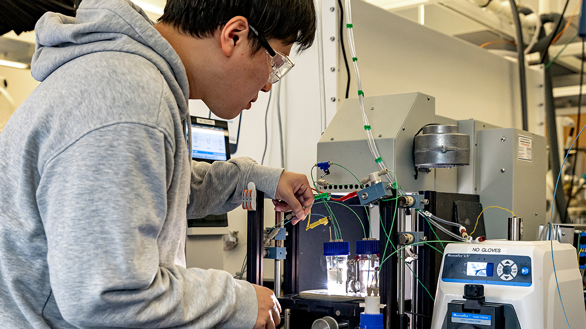
Carolina assistant professor Stephen Smith uses a microscope he designed himself in order to know for certain what neurons — and which kind of neurons — fire as a result of specific stimuli.
Spencer Smith peers through a microscope of his own creation, trying to home in on a single neuron.
The cell is tiny, about 0.015 millimeters in diameter. He attempts to clamp a microscopic pipette directly onto the neuron’s dendrite — an arm-like extension of the cell. He wants to explore its function in greater detail than anyone has ever done. It’s tedious work that requires otherworldly patience, a lot of time, and a powerful microscope. Finally, Smith manages to connect the pipette. Then, he listens and he looks.
Eventually, he witnesses a spike of current. The dendrite in the brain of a live mouse has fired an independent electrical signal. That is, the signal wasn’t created elsewhere and then appeared in the dendrite.
To a neuroscientist, this is big news. To anyone interested in how the brain works, this should be surprising news. Scientists had long thought that dendrites were merely the passive circuits that transmit electrical signals from synapse to the main cell bodies of neurons. Smith’s research shows that when a mammal is making sense of visual information, dendrites can actually create the signals to process that information themselves. Turns out, dendrites aren’t just wiring in the brain — which scientists had thought for decades. Dendrites are like mini-neural computers.
“Imagine you’re reverse engineering a piece of alien technology, and what you thought was simple wiring turns out to be transistors that compute information, said Smith, an assistant professor of cell biology and physiology and member of the UNC Neuroscience Center. “That’s what this finding is like.”
Except we’re talking about the mammalian brain. “The implications are exciting to think about,” he said.
Read more here, http://news.unchealthcare.org/news/2014/august/the-two-photon-future